
Lubusky M., Prochazka M., Dhaifalah I., Horak D., Geierova M. Santavy J. Fetal enterolithiasis: prenatal sonographic and MRI diagnosis in two cases of urorectal septum malformation (URSM) sequence. Prenat. Diagn., 2006, 26 (4), p. 345-349. (IF-1,514)
ABSTRACT
Objectives: Enterolithiasis (multiple calcifications of intraluminal meconium) is a rare, prenatal ultrasonographic finding. In this study, our aim was to evaluate the prenatal diagnostic features and discuss the management of the patients.
Methods: The data of two cases of prenatally diagnosed fetal enterolithiasis were collected from ultrasound scan, magnetic resonance imaging (MRI) and neonatal or postnatal autopsy records. The findings were evaluated in both prenatal and postnatal periods. Chromosomal analysis was performed in one case. An evaluation of primary and secondary malformations was done. Coexisting anomalies were searched for via radiology, neonatal surgery and histopathology.
Results: Malformations in two cases (both males) with partial and complete urorectal septum malformation (URSM) sequence were described. The absence of an anal opening and presence of a fistula between the urinary and gastrointestinal tract were common findings. These features were considered as primary malformations contributing to the formation of enterolithiasis. Secondary anomalies (urinary and gastrointestinal system malformations, pulmonary hypoplasia, genital and other coexisting anomalies) were evaluated.
Conclusions: The prenatal detection of enterolithiasis carries a poor prognosis.Most of the previously reported cases were invariably associated with major fetal malformations of the urinary and gastrointestinal tract. It is a warning sign for large bowel obstruction with or without enterourinary fistula. Therefore, adequate gastrointestinal and urologic studies must be undertaken after birth for the final diagnosis. There is a high mortality rate in the reported cases, mostly attributed to associated anomalies, and all survivors required neonatal surgery. It is important to differentiate the partial from the full URSM sequence because the prognosis in the partial URSM sequence is generally good, with long-term survival being common. Copyright 2006 John Wiley & Sons, Ltd.
Key words: fetal enterolithiasis; urorectal septum malformation sequence


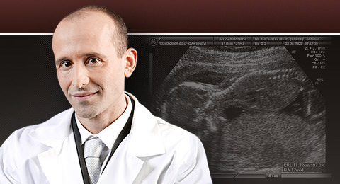
Contact
Professor Marek Lubusky, MD, PhD, MHA
THE FETAL MEDICINE CENTRE
Department of Obstetrics and Gynecology
Palacky University Olomouc, Faculty of Medicine and Dentistry
University Hospital Olomouc
Zdravotníků 248/7, 779 00 Olomouc, Czech Republic
Tel: +420 585 852 785
Mobil: +420 606 220 644
E-mail: marek@lubusky.com
Web: www.lubusky.com