
Lubusky M., Prochazka M., Dhaifalah I., Horak D., Geierova M. Santavy J. Fetal enterolithiasis: prenatal sonographic and MRI diagnosis in two cases of urorectal septum malformation (URSM) sequence. Prenat. Diagn., 2006, 26 (4), p. 345-349. (IF-1,514)
Introduction
The finding of extraluminal (intra-abdominal) calcifications is commonly reported and usually indicates intrauterine intestinal perforation with intraperitoneal extravasation of meconium (meconium peritonitis), most often associated with intestinal obstruction and/or atresias (Kamensky and Howard, 1952). Intraluminal calcification of meconium is more rare and mostly appears to result from the mixing of stagnant urine and meconium in utero. The presence of the intraluminal calcifications in a dilated loop of intestine, particularly with an associated urinary tract abnormality, should suggest a fistula between the urinary and the gastrointestinal tracts.
Multiple foci of intraluminal calcified meconium, also known as enterolithiasis, has been previously described as a radiologic finding in newborn infants with several pathologic conditions, including imperforate anus (Berdon et al., 1975; Felman et al., 1975; Cook, 1978; Selke and Cowley, 1978; Berger and Bar-Maor, 1980; Pouillaude et al., 1987; Anderson et al., 1988), persistent cloaca (Bear and Gilsanz, 1981), multiple gastrointestinal atresias (Martin et al., 1976; Daneman and Martin, 1979; Yousefzadeh et al., 1984), small bowel stenosis (Camp and Roberts, 1949), total colonic aganglionosis (Hirschsprung disease) (Fletcher and Yullish, 1978), and functional ileal obstruction (Rickham, 1957). The prenatal identification of enterolithiasis therefore is an important obstetrical goal because the existence of underlying gastrointestinal pathology, the presence of associated urogenital anomalies and the need of neonatal surgery can be anticipated.
In our reports we present two cases of enterolithiasis in the fetuses, one with partial and the other complete urorectal septum malformation (URSM) sequence, which were detected prenatally by ultrasound scan and magnetic resonance imaging (MRI).
Case 1
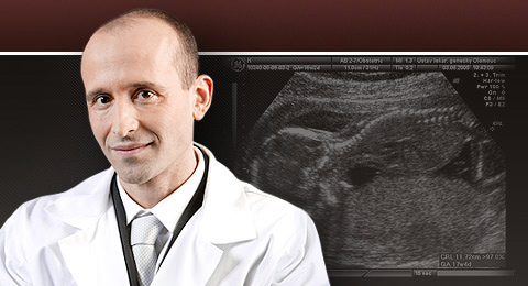
The mother, a 19-year-old Caucasian woman, gravida 1, para 0, was referred to the ultrasound unit of the department of medical genetic and fetal medicine at 18 weeks of spontaneous pregnancy for routine screening. There was no family history of congenital malformations. On sonographic examination, a monochorionic, diamniotic twin pregnancy was diagnosed. Twin A had dilated distal bowel loops with enterolithiasis (an ultrasonographic examination revealed en echogenic bowel with multiple foci of calcified meconium intraluminally) (Figure 1). Twin B had dilated distal bowel loops. A possible diagnosis of anal atresia was made. The genitalia could not be clearly determined. In view of the abnormal sonographic findings, amniocentesis was performed. Both fetuses had a normal 46,XY karyotype. The patient was referred to the high-risk clinic. Fetal biometry was appropriate for gestational age and a normal amount of amniotic fluid was observed. To further rule out anorectal malformation MRI was performed and demonstrated dilated distal bowel loops (with enterolithiasis in twin A) (Figure 2). The parenchyma of the kidneys and urinary bladder appeared normal. Pregnancy was terminated by caesarean section at 35 weeks of gestation after premature rupture of the membranes. Twin A was a male weighing 1950 g. Twin B weighed 2350 g. Post-partum examination revealed close placental insertions of both umbilical cords, each containing three vessels. Over a length of 25 cm the umbilical cords, separated by amniotic membranes, ran in such close proximity that they appeared to have a common course. The monochorionic, diamniotic twin pregnancy was certified and concordant fetal abnormalities were diagnosed. Both neonates had a single perineal opening that drained a common cloaca in combination with anal atresia. The cloaca drained the bladder and colon separately. There was a short and hypoplastic colon, dilated distal bowel loops (in the twin A with enterolithiasis) and a fistula between the colon and the bladder. External genital malformations included: cloaca with a single opening, perineal hypospadias, bifid scrotum and penoscrotal transposition. There were also pelvic and sacral abnormalities. The diagnosis of partial URSM sequence was confirmed postnatally and the babies underwent corrective urogenital and intestinal surgeries. At present they are 2 years of age and their health condition is generally good.
Case 2
The mother, a 28-year-old Caucasian woman, gravida 2, para 1, presented to the ultrasound unit of the department of medical genetic and fetal medicine at 20 weeks of a spontaneous pregnancy, because of oligohydramnios. On sonographic examination, fetal biometry was appropriate for gestational age and a severe oligohydramnios to anhydramnios was observed. The fetus had dilated bowel loops with enterolithiasis (an ultrasonographic examination revealed multiple echogenic foci of calcified meconium intraluminally) (Figure 3). There was right hydronephrosis (renal pelvis dilatation in anteroposterioric diameter up to 10 mm, with no parenchyma reduction or calices distention), as well as a dilated ureter up to 8 mm. The parenchyma of the left kidney appeared normal without ureter distention. The urinary bladder was not visible and the genitalia could not be clearly determined. A possible diagnosis of complete URSM sequence was made. MRI was performed and demonstrated dilated bowel loops with enterolithiasis; however, the urinary bladder remained undetectable (Figure 4). Because of the parents’ demand, the pregnancy was terminated. The diagnosis of complete URSM sequence was confirmed postnatally. External examination showed ambiguous (unrecognizable) genitalia, no phallic structure and no perineal or anal opening (blank perineum). On autopsy, pulmonary hypoplasia was not described. Autopsy showed abdominal testes, distention of the urinary bladder (35 × 15 mm) with outlet obstruction, a fistula between the dilated distal small intestine (terminal ileum) and the bladder, distal colon and rectal atresia. The colon ended in a dilated meconium-filled, blind pouch. Intraluminal calcification of meconium was described in the dilated colon and the bladder. There was right hydronephrosis (dilatation of the renal pelvis without parenchyma reduction and distention of calices) and a dilated ureter without distinct connection to the bladder. The parenchyma of the left kidney was normal and the ureter typically drained into the bladder. Dysplastic changes in both kidneys were not described. The diagnosis of a megacystis with enterolithiasis was set postnatally according to the post-mortem examination. Chromosome analyses were not done.
Discussion
These reports document the prenatally diagnosed cases of enterolithiasis in the fetuses with partial and complete URSM sequences. The URSM sequence consists of the absence of the perineal and anal opening in association with ambiguous genitalia and urogenital, colonic and lumbosacral anomalies. The abnormalities of this condition are thought to arise early in development from incomplete subdivision of the primitive cloaca and lack of breakdown of the cloacal membrane (Escobar et al., 1987; Wheeler et al., 1997). A less severe form of the URSM sequence is referred as the partial URSM sequence (Wheeler and Weaver, 2001). Individuals with a partial URSM sequence typically have a single perineal/anal opening that serves as an outlet for a common cloaca and conduit for urine and feces to the outside. The full URSM sequence is usually lethal in the newborn period owing to pulmonary hypoplasia resulting from severe oligohydramnios. In our Case 2, pulmonary hypoplasia was not described. The pregnancy was terminated at 21 weeks and it is likely that pulmonary hypoplasia resulting from severe oligohydramnios could have developed later. It is important to differentiate the partial from the full URSM sequence because the prognosis in the partial URSM sequence is generally good, with long-term survival being common.
The mechanism by which intraluminal meconium calcifies is poorly understood. It has been suggested that both the prolonged statis of the meconium and the interaction between urine and meconium play significant roles (Berdon et al., 1975; Anderson et al., 1988; Miller et al., 1988; Sepuvelda et al., 1994). The meconium, being made of decimated products from the bowel wall and also from the bile salts that are concentrated because of enterohepatic circulation, is high in calcium. It is probable that a change of pH causes precipitation of the calcium salt. The calcifications lie in the centre of the small balls of meconium. Presumably the calcium is precipitated and the urine, with the bowel movements, causes the meconium to be rolled into ball-like configurations around the aggregates of calcium. However, the relative contribution of the urine-meconium mixture has not yet been elucidated. In the cases of lower bowel obstruction such as imperforate anus, enterolithiasis generally is associated with the presence of urorectal fistula, suggesting that the presence of urine in the fetal bowel plays a critical role in the process of meconium calcification (Berdon et al., 1975; Anderson et al., 1988; Miller et al., 1988; Sepuvelda et al., 1994). However, evidences suggest a limited role for urine-meconium mixture in the formation of calcified enteroliths: (1) the vast majority of neonates with imperforate anus and rectourinary fistula have no enterolithiasis demonstrated by preoperative radiologic evaluations, (2) several cases of enterolithiasis in newborn infants with imperforate anus but without demonstrable rectourinary fistula at surgery or autopsy have been reported in the literature (Berdon et al., 1975; Berger and Bar-Maor, 1980; Pouillaude et al., 1987; Sepuvelda et al., 1994), (3) enterolithiasis also may occur in neonates with small bowel obstruction and no associated fistula (Camp and Roberts, 1949; Martin et al., 1976; Dodat et al., 1983), (4) it has, however, been previously described in Hirschsprung disease (Fletcher and Yullish, 1978), (5) in adults, enterolithiasis occurs exclusively in the cases in whom the pathologic findings indicate local stasis of fecal material (Paige et al., 1987) and (6) in several cases the authors have argued that although in normal fetuses the mixture of meconium and urine in the fetal bowel is a physiologic phenomenon, intraluminal calcification does not occur (Yousefzadeh et al., 1984; Pouillaude et al., 1987; Sheth, 1993). The last of these arguments may be taken as evidence that local pH may play a significant role. Although fetal urine is alkaline, passage through the stomach changes its pH to an acidic value owing to mixture with the gastric secretions. Therefore, in normal circumstances the urine reaching the fetal bowel is acidic and not alkaline as in the cases of a fistula between the urinary and gastrointestinal tract. In addition, it is noteworthy that in almost half of the neonates in whom enterolithiasis was associated with imperforate anus, esophageal atresia was also present (Berdon et al., 1975; Anderson et al., 1988; Simma et al., 1992; Duncan, 2001). This provides further evidence that local pH is important, as in these cases the normal acid contribution into the fetal bowel content is prevented by the upper gastrointestinal obstruction. A similar situation is caused by anhydramnios in the case of complete URSM. We have no clear explanation why of both twins (our Case 1) with concordant malformations only one twin developed enterolithiasis. In our opinion it could be because of a different degree of stasis of the meconium of the twins and the interaction between urine and meconium, which would have altered the pH and thus may have facilitated the development of enterolithiasis.
Enterolithiasis is a rare, prenatal ultrasonographic finding. Most of the previously reported cases were invariably associated with major fetal malformations. Of the nine cases diagnosed prenatally (Shalev et al., 1983; Grant et al., 1990; Mandell et al., 1992; Simma et al., 1992; Sepuvelda et al., 1994; Achiron et al., 1998, 2000), six had imperforate anus, one had a stenotic ectopic anus and one had an anorectal agenesis (Mandell et al., 1992; Sepuvelda et al., 1994). Six cases had fistula between the anorectum and the urinary tract, and in eight cases genito-urinary malformations coexisted resulting in severe oligohydramnios in two (Simma et al., 1992; Sepuvelda et al., 1994). Achiron et al. described a case of fetal anhydramnios and enterolithiasis in an anatomically normal fetus that on post-mortem examination was found to have end-stage liver disease (fetal hepatorenal syndrome) (Achiron et al., 1998). The enterolithiasis in this case may have resulted from abnormal bile secretion due to the liver disease and decreased bowel fluid due to the anhydramnios. This case also questions the hypothesis that a mixture of urine and meconium, through an anomalous urino-intestinal connection, is necessary for the formation of enterolithiasis.
Although previously it may have been difficult to determine whether calcifications were intraluminal or extraluminal, current high-resolution sonographic and MRI equipments provide better anatomic detail and facilitates the evaluation of fetal intestinal anomalies. The prenatal diagnosis (detection) of enterolithiasis carries a poor prognosis (Anderson et al., 1988; Sepuvelda et al., 1994). It is a warning sign for large bowel obstruction with or without enterourinary fistula. Therefore, adequate gastrointestinal and urologic studies must be undertaken after birth for the final diagnosis. There is a high mortality rate in the reported cases, mostly attributed to associated anomalies, and all the survivors required neonatal surgery.
Acknowledgements
This study was supported by the Medical Faculty of Palack´y University Olomouc ‘Safety of Ultrasound in Medicine’.
References
- Achiron R, Daniel Y, Levran D, Lipitz S. 1998. Fetal enterolithiasis and anhydramnios; due to in utero hepatorenal syndrome? Prenat Diagn 18: 1195–1197.
- Achiron R, Frydman M, Lipitz S, Zalel Y. 2000. Urorectal septum malformation sequence: prenatal sonographic diagnosis in two sets of discordant twins. Ultrasound Obstet Gynecol 16: 571–574.
- Anderson S, Savader B, Barnes J, Savader S. 1988. Enterolithiasis with imperforate anus. Report of two cases with sonographic demonstration and occurrence in a female. Pediatr Radiol 18: 130–133.
- Bear JW, Gilsanz V. 1981. Calcified meconium and persistent cloaca. AJR Am J Roentgenol 137: 867–868.
- Berdon WE, Baker DH, Wigger HJ, et al. 1975. Calcified intraluminal meconium in newborn males with imperforate anus: enterolithiasis in the newborn. Am J Roentgenol Radium Ther Nucl Med 125: 449–455.
- Berger J, Bar-Maor JA. 1980. Intraluminal intestinal calcifications in a newborn with atresia of the esophagus and imperforate anus. Clin Pediatr 19: 770–772.
- Camp R, Roberts MH. 1949. Multiple calcareous deposits in the intestinal tract of the newborn. Report of a case associated with stenosis of the ileum. Am J Dis Child 78: 393–395.
- Cook PL. 1978. Calcified meconium in the newborn. Clin Radiol 29: 541–546.
- Daneman A, Martin DJ. 1979. A syndrome of multiple gastrointestinal atresias with intraluminal calcification. Pediatr Radiol 8: 227–231.
- Dodat H, Galifer B, Grisard M, Robert M, Sauvage P. 1983. Neonatal enterolithiasis (or intraluminal meconial calcifications): apropos of four cases. Chir Pediatr 24: 183–185.
- Duncan AW. 2001. Quiz case. Multiple calcification of intraluminal meconium enterolithiasis. Eur J Radiol 37: 120–122.
- Escobar LF, Weaver DD, Bixler D, Hodes ME, Mitchel M. 1987. Urorectal septum malformation sequence: report of six cases and embryological analysis. Am J Dis Child 141: 1021–1024.
- Felman AH, Walker RD, Donelly WH, Gerami S. 1975. Supralevator imperforate anus with unusual associated anomalies: colonic ureteral ectopy, intraluminal calcified meconium. Pediatr Radiol 3: 78–80.
- Fletcher BD, Yullish BS. 1978. Intraluminal calcifications in the small bowel of newborn infants with total colonic aganglionosis. Radiology 126: 451–455.
- Grant T, Newman M, Gould R, Schey W, Perry R, Brandz T. 1990. Intraluminal colonic calcifications associated with anorectal atresia. Prenatal sonographic detection. J Ultrasound Med 9: 411–413.
Pozn.: Tabulky, grafy a obrázky naleznete v souboru Format PDF ».

Contact
Professor Marek Lubusky, MD, PhD, MHA
THE FETAL MEDICINE CENTRE
Department of Obstetrics and Gynecology
Palacky University Olomouc, Faculty of Medicine and Dentistry
University Hospital Olomouc
Zdravotníků 248/7, 779 00 Olomouc, Czech Republic
Tel: +420 585 852 785
Mobil: +420 606 220 644
E-mail: marek@lubusky.com
Web: www.lubusky.com